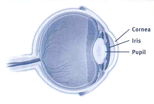
Corneal topography is a computer-assisted procedure used to measure the curvature of the cornea, the clear front window of the eye. The corneal topographer projects illuminated circles on the cornea that are reflected back to the computer and used to produce a map of the cornea. This map can reveal any irregularities in the cornea’s curvature. Other devices measure the corneal elevation in three dimensions, and these measurements are converted into a corneal curvature map.

Normal Eye Anatomy
Corneal topography is commonly used to help follow the progression of keratoconus and to assist in fitting patients with contact lenses to treat the visual distortions caused by this condition. It is also one tool that may be used to help with certain refractive procedures such as LASIK. Following corneal transplant surgery, corneal topography helps the surgeon identify where to selectively remove sutures to smooth the shape of the new cornea.
Corneal topography is quick and painless. A technician will ask you to sit comfortably and rest your head against a bar on the topographer. You then look into a lighted bowl. The technician takes a picture, which the computer uses to analyze the curvature of the cornea and to produce an image that the Advanced Eye Center ophthalmologist or optometrist will use in your treatment.


